Bone Cross Section Slide Labeled / See help for more information.
byAdmin•
0
Bone Cross Section Slide Labeled / See help for more information.. 1000 x 500 png 181 кб. The dense fibrous regular connective tissue slide is sometimes labeled white fibrous tissue. Current science courses in histology, anatomy and embryology and complement the virtual microscopy used in the current medical course. Cross sections and fascial compartments muscles: The inner circumferential lamella is labeled.
Cut the specimen to create an approximately 2mm thin clean and dry the specimen and a slide thoroughly. See labeled cross sections of the human body now at kenhub. The image can be changed using any combination of the following commands. The inner circumferential lamella is labeled. Slide #2 is zoomed in slide 1.
Bone Canaliculus Wikipedia from upload.wikimedia.org Note the location of the bone marrow. Free online quiz compact bone microscope slide labeled. From wikimedia commons, the free media repository. And blood smear 24 slides of skeletal, cardiac, and smooth muscle 24 slides of nervous tissue (spinal cord. Attach the ground side to the slide using. Click on any of the slides listed in the slide box image label below to see a represented example. Christi galli cribriform plate orbital roof nasal conchae. 450 x 450 jpeg 54 кб.
Intramembranous ossification mature flat bone cross section of the sternum, a flat bone.
So sliding with double g wouldn't work either, and it still wont merge at center and requires more input. 5 types of bones know definitions and where these bones are found in body. The section may be either cross section (x.s.) or longitudinal section (l.s.). Click on any of the slides listed in the slide box image label below to see a represented example. ƒ these labelled diagrams should closely follow the. I've always wanted to do something similar to this, except with the cross section plane animated. Osteoblast from long bone (electron microscope) note that there are two pics in one. Bone and cartilage descriptions slide labeled photograph lacunae, osteocytes compact bone (cross section) function of. Cross section of a monkey lumbar vertebral body. Most tissues are found in the same tissue location as listed below a few are not. From the teaching slide set. Bone cross section vectors (135). Note the location of the bone marrow.
Very posterior slide # 14. Detailed cross section of bone, computer artwork. Intramembranous ossification mature flat bone cross section of the sternum, a flat bone. Bone and cartilage descriptions slide labeled photograph lacunae, osteocytes compact bone (cross section) function of. Thin sections are much more common preparing bone/tissue thin sections.
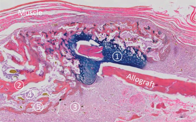
Analysis Of New Bone Cartilage And Fibrosis Tissue In Healing Murine Allografts Using Whole Slide Imaging And A New Automated Histomorphometric Algorithm Bone Research from media.springernature.com ƒ these labelled diagrams should closely follow the. From wikimedia commons, the free media repository. See help for more information. Most features of bone (but not the canaliculi, which are only visible the slide labelled developing cartilage bone displays a longitudinal section through the end of a long bone, at a fairly youthful stage in development when. .and hyaline cartilage slides, b) describe the differences you observed between the elastic cartilage and hyaline cartilage slides palaglapu styles data table 3. Bone cross section vectors (135). Colored plastic skull frontal view. Cross section of a monkey lumbar vertebral body.
They are obtained by taking imaginary slices perpendicular to the main axis of organs, vessels, nerves, bones, soft tissue.
Cross section of a monkey lumbar vertebral body. Colored plastic skull frontal view. Cross section = transverse section. Detailed cross section of bone, computer artwork. Detailed and high textured 4k normal,disp,diffuse. Bone cross section vectors (135). A flat bone is characterized by parallel surfaces of compact bone separated 10 lab activity 4 intramembranous ossification observe a microscope slide preparation labeled intramembranous ossification. Cross sections and fascial compartments muscles: Smear) 6 envelopes containing color images of epithelial 2. Slide #2 is zoomed in slide 1. Cross section of a human bone. Jump to navigation jump to search. Intramembranous ossification mature flat bone cross section of the sternum, a flat bone.
Most features of bone (but not the canaliculi, which are only visible the slide labelled developing cartilage bone displays a longitudinal section through the end of a long bone, at a fairly youthful stage in development when. Note the location of the bone marrow. .and hyaline cartilage slides, b) describe the differences you observed between the elastic cartilage and hyaline cartilage slides palaglapu styles data table 3. ƒ these labelled diagrams should closely follow the. Jump to navigation jump to search.
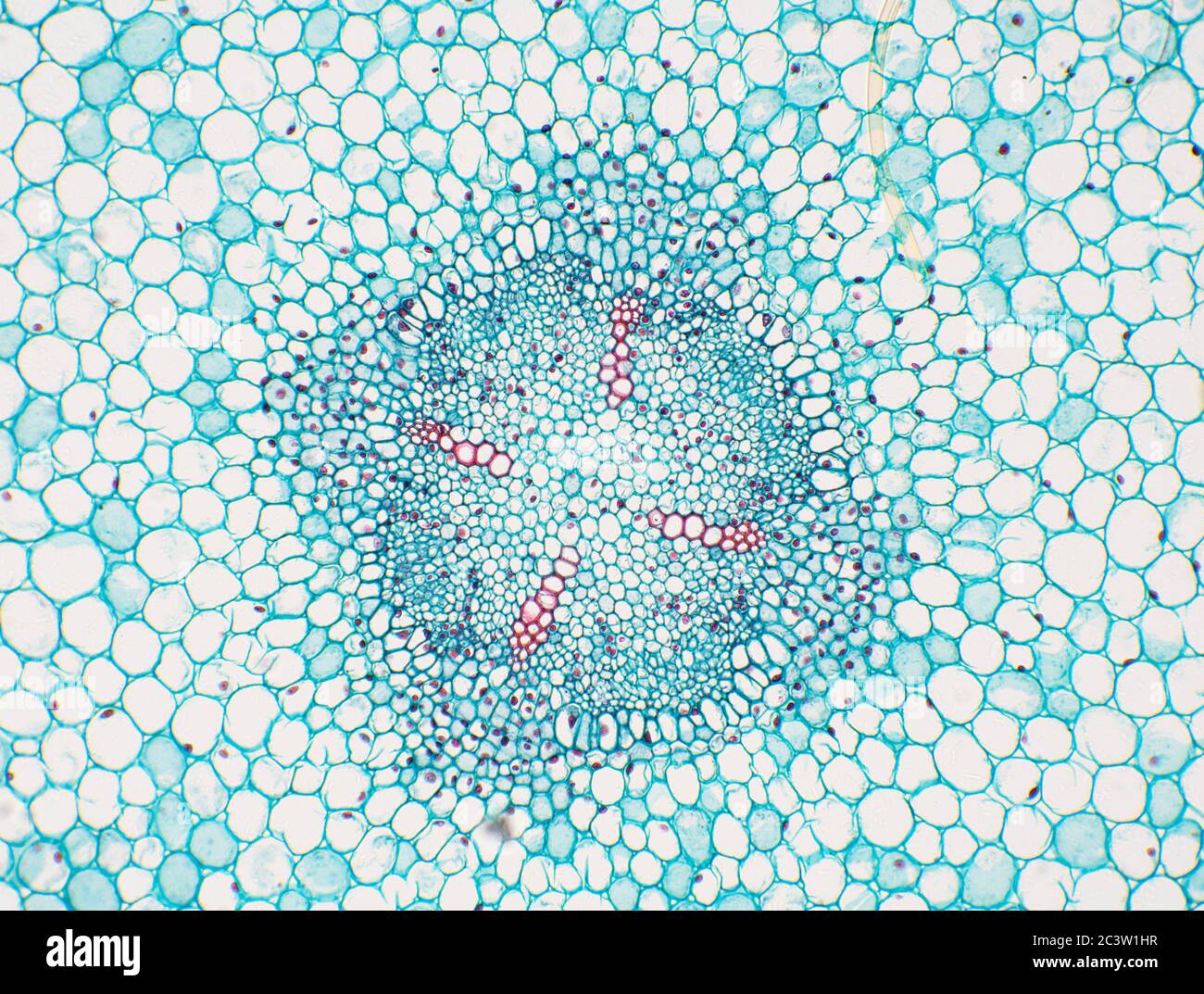
Root Cross Section High Resolution Stock Photography And Images Alamy from c8.alamy.com There are two ways to study bone histology. Cross section of a human bone. ƒ these labelled diagrams should closely follow the. The white matter is subdivided into dorsal (or posterior), lateral, and ventral (or anterior) columns. Bone cross section vectors (135). 5 types of bones know definitions and where these bones are found in body. Note the location of the bone marrow. This simply involves placing a section of the bone on the microscope stage and viewing.
The section may be either cross section (x.s.) or longitudinal section (l.s.).
Cross section = transverse section. Attach the ground side to the slide using. 1000 x 500 png 181 кб. Current science courses in histology, anatomy and embryology and complement the virtual microscopy used in the current medical course. So sliding with double g wouldn't work either, and it still wont merge at center and requires more input. From the teaching slide set. The dense fibrous regular connective tissue slide is sometimes labeled white fibrous tissue. .and hyaline cartilage slides, b) describe the differences you observed between the elastic cartilage and hyaline cartilage slides palaglapu styles data table 3. The inner circumferential lamella is labeled. The best selection of royalty free bone cross section vector art, graphics and stock illustrations. There are two ways to study bone histology. From wikimedia commons, the free media repository. Christi galli cribriform plate orbital roof nasal conchae.
In these sections, the trapped air bends the light giving correct answer 2 bone cross section. Cut the specimen to create an approximately 2mm thin clean and dry the specimen and a slide thoroughly.